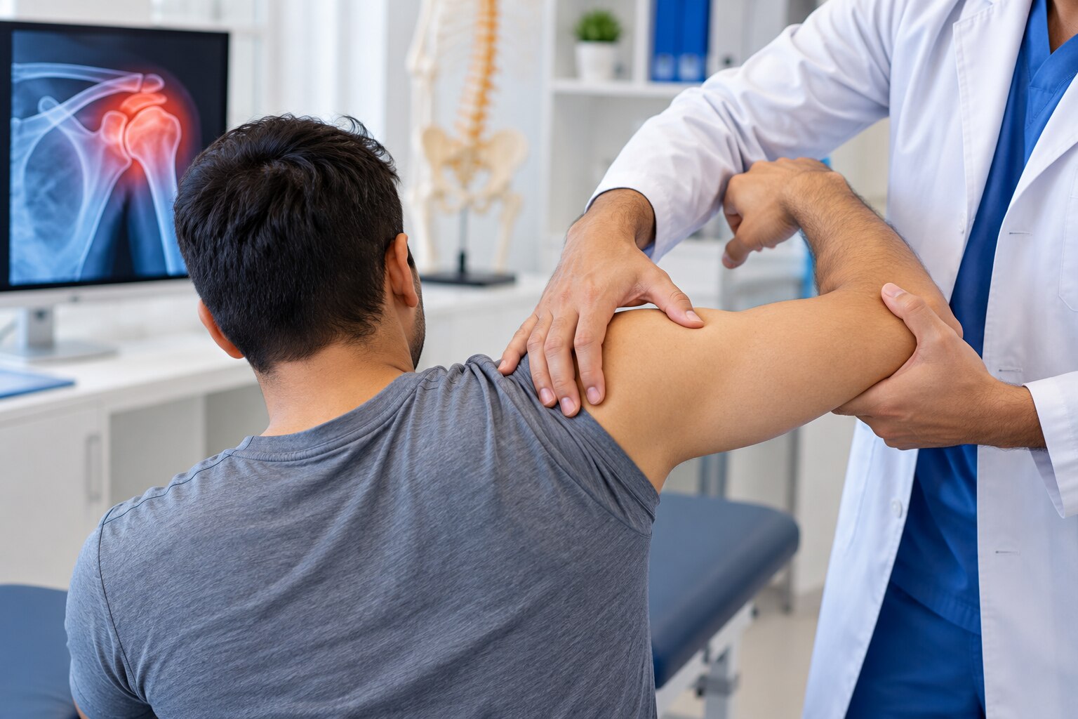
A suspected dislocation needs more than a joint being placed back into position: the clinician must check for fractures, nerve damage and impaired blood flow before and after treatment. You will learn how to protect the injury, recognise when to call an ambulance, understand the examination and imaging process, and judge whether local care is equipped for your situation.
Key takeaways
- Support the joint as found and never try to reduce it yourself.
- Expect a physical examination plus X-rays to confirm the dislocation and check for fractures.
- Reduction restores alignment; sedation or pain relief helps control discomfort during the procedure.
- Choose a clinic with urgent imaging, reduction facilities and follow-up rehabilitation.
What should you do before reaching medical care?
Keep the joint supported in the position you found it and arrange urgent medical assessment; do not try to put it back yourself.
1. Steady the injured limb with clothing, cushions or a splint without forcing it straight. Remove rings, watches and tight jewellery before swelling increases.
2. Apply a cold pack wrapped in cloth for 10–15 minutes at a time. Never place ice directly on the skin.
3. Do not pull, straighten, massage or otherwise manipulate the joint. Do not ask an untrained person to reduce it; force can worsen a fracture or damage a nerve or blood vessel.
4. Give no food or drink when reduction, sedation or surgery could be needed. Keep the person still and take any prescribed medicines or allergy information to the clinician.
5. Call an ambulance rather than using private transport for a cold, pale or blue limb; an absent pulse; numbness or weakness; uncontrolled bleeding; an open wound; severe neck or back pain; loss of consciousness; suspected hip dislocation after major trauma; or severe pain after a high-energy impact.
A stable-looking deformity still needs urgent assessment. A fracture, nerve injury or blood-vessel injury can sit behind it, even when the person can move the limb. An orthopedic surgeon in Hinjewadi for dislocation will need to assess the joint safely rather than rely on appearance alone.
How will the surgeon identify the joint injury?
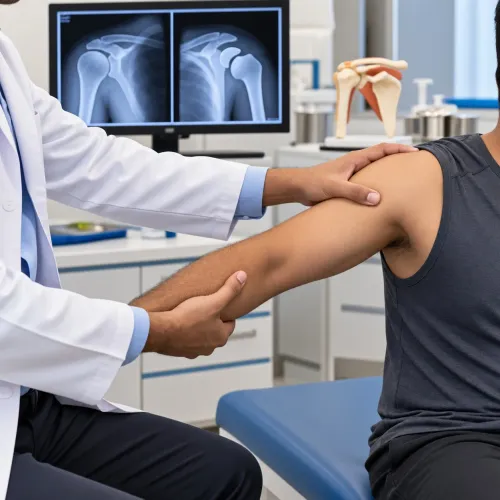
Assessment starts by identifying the joint, direction of displacement, cause and possible fracture. A complete dislocation means the joint surfaces lose contact; a subluxation means they partly separate.
The orthopedic doctor hinjewadi patients consult will ask which movement or impact caused it, the exact time, whether it dislocated before and whether it returned to position on its own.
The orthopedic surgeon hinjewadi patients seek will examine and document:
- Skin breaks, swelling, deformity and tenderness
- Pulse and capillary refill
- Sensation and motor function
Recordings are repeated after reduction. A joint that appears to have slipped back still needs examination because cartilage, ligaments, tendons, nerves or bone may be injured.
| Joint | Clue during assessment | Why it matters |
|---|---|---|
| Shoulder | Squared-off contour | Suggests the ball has left the socket |
| Elbow | Visible bending and swelling | Fracture or ligament damage may accompany it |
| Finger | Shortening or rotation | Indicates abnormal alignment |
| Kneecap | Shift towards one side | Suggests patellar displacement |
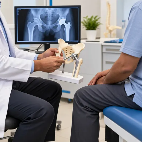
| Hip | Shortened leg turned inward or outward after major trauma | Requires emergency assessment |
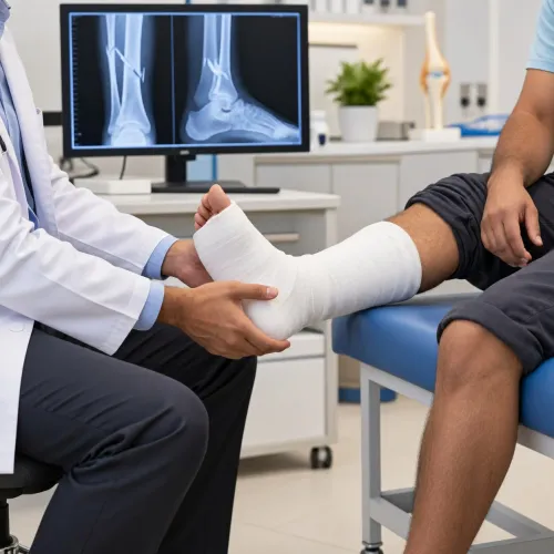
| Ankle | Deformity, swelling or inability to bear weight | Fracture-dislocation must be excluded |
| Jaw | Inability to close the mouth normally | Indicates possible jaw dislocation |
What imaging and reduction treatment should you expect?
Dislocation treatment hinjewadi is not one standard procedure. The surgeon identifies the joint, displacement direction, injury mechanism and presence of a fracture before selecting imaging and reduction.
| Modality | What it shows | When it is used |
|---|---|---|
| Plain X-rays | Alignment and fractures | Before reduction when the mechanism, deformity or examination raises fracture concern; extra views can reveal a hidden break |
| Post-reduction X-rays | Joint position and obscured injury | After reduction to confirm alignment and detect a fracture hidden earlier |
| CT | Complex bone injury | For a fracture-dislocation or detailed assessment of broken joint surfaces |
| MRI | Labral, ligament, tendon and cartilage injury | When these findings will change treatment, particularly after the acute injury is stabilised |
- The surgeon distinguishes a simple dislocation from a fracture-dislocation, growth-plate injury, nerve injury and blood-vessel injury. Reducing a joint without imaging can be unsafe when a fracture is suspected or the mechanism is unclear.
- Reduction is commonly performed with pain relief, procedural sedation or another monitored muscle-relaxing analgesic technique. The choice depends on the joint, your health, injury complexity and available monitoring.
- After sedation, expect observation until you are alert and stable. Arrange for someone else to take you home; do not drive yourself.
- Once alignment is restored, the clinician rechecks pulse, capillary refill, sensation and movement. A normal-looking joint does not replace this neurovascular check.
What happens after the joint is put back?
A successful reduction is only the midpoint of treatment. The surgeon takes new X-rays when indicated, confirms that the joint is aligned, checks pulse, capillary refill, sensation and movement, and looks for fractures hidden before reduction.
A sling, splint or another support protects the joint when needed, but there is no safe universal sling duration. Ask exactly when to remove it and begin range-of-motion exercises. Prolonged shoulder immobilisation can cause stiffness, particularly in older adults.
Your follow-up plan depends on the joint and your risk of recurrence:
- After a first anterior shoulder dislocation, younger athletes have a higher recurrence risk than older adults. Repeated minor slipping episodes need an instability assessment rather than repeated unsupervised reductions.
- Elbow dislocations need follow-up because stiffness and lost movement can develop. If the joint is stable, supervised early movement may begin when the surgeon considers it safe.
- Surgery is not automatic after a first uncomplicated dislocation. It becomes more likely when a fracture needs fixation, instability persists, a major tendon or labral injury is present, dislocations recur, or your work or sport makes recurrence unacceptable.
A hip dislocation remains an emergency. Reduction is performed as soon as safely possible, often within about six hours, because delay increases the risk of femoral-head osteonecrosis.
How should you choose an orthopedic clinic in Hinjewadi?
Choose an orthopedic clinic Hinjewadi residents can reach, but confirm its limits before booking. An outpatient consultation does not prove that the clinic can manage every dislocation safely.
| Option | Ask or expect | Choose it when |
|---|---|---|
| Clinic assessment | Same-day radiographs, pulse and nerve checks, and documented findings before and after reduction | The injury is non-critical and the limb is warm, coloured and sensitive |
| Hospital-supported care | Monitored sedation at the clinic or through a hospital, with observation afterwards | Reduction requires pain control or muscle relaxation |
| Emergency or surgical facility | Transfer for complex fracture-dislocations, open injuries or vascular damage | Imaging or examination shows risks beyond outpatient care |
Ask who will provide emergency follow-up if pain, deformity, numbness, increasing swelling or loss of movement returns after reduction. Confirm who reassesses alignment, circulation, nerve function and associated fractures, and who arranges the sling, splint or rehabilitation plan.
For a non-critical injury, Latkar Multi-speciality Clinic can be part of that local assessment pathway; the treating orthopedic surgeon decides whether clinic care, hospital treatment or surgery is appropriate. Verify an Indian orthopaedic doctor’s registration in the National Medical Commission’s Indian Medical Register.
Take previous imaging, medication and allergy details, plus the injury timeline. Choose emergency services instead of a routine appointment with an orthopedic surgeon Hinjewadi patients may book if the limb is pale, cold, blue, numb or weak, or if there is an open wound, suspected hip dislocation, severe trauma or possible fracture.
Related product
 | Bone Fractures & Joint Dislocation DISLOCATION Bone Fractures & Joint Dislocation Two of the most common bone and joint injuries are fractures and dislocations. |
Frequently asked questions
What should you do before reaching medical care for a dislocation?
Keep the joint supported in the position you found it, avoid movement, apply a wrapped cold pack, and arrange urgent medical assessment. Do not try to put the joint back yourself.
How does an orthopedic surgeon identify a dislocated joint?
The surgeon checks the injury’s appearance, pain, movement, skin condition, circulation and nerve function. They also ask how the injury happened and use imaging to confirm the diagnosis.
What imaging and treatment should you expect for a dislocation?
X-rays usually confirm the joint position and show fractures. The surgeon then reduces the joint using controlled techniques, with pain relief or sedation when appropriate, and repeats imaging if needed.
What happens after the joint is put back?
The team checks circulation, sensation and movement again, provides a sling, splint or brace when needed, and gives instructions for pain control, activity and follow-up physiotherapy.
How should you choose an orthopedic clinic in Hinjewadi?
Choose an orthopedic clinic with urgent assessment, on-site or promptly available X-rays, trained staff for reduction, emergency referral access and a clear follow-up rehabilitation plan.
Keywords