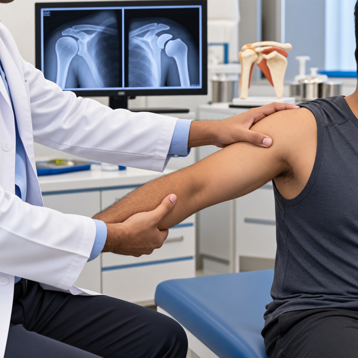
A dislocated joint needs more than a quick visual check: the clinician must assess nerves, blood flow, associated fractures and the safest way to restore alignment. You will learn how to respond before reaching care, what happens during reduction, when a hospital is safer than a nearby clinic and what recovery involves.
Key takeaways
- Keep the joint still and support it; do not attempt to push it back yourself.
- Seek emergency care for numbness, pale or cold skin, open wounds, or severe deformity.
- X-rays check alignment and fractures before and after joint reduction.
- Follow the rehabilitation plan to restore movement, strength and joint stability.
How to recognise a dislocation and protect the joint before care
A dislocation means the joint surfaces have lost their normal alignment: one bone end has been forced out of its socket. It may look visibly deformed and cause severe pain, swelling and inability to move the limb. Bone fractures break bone; sprains stretch or tear ligaments; tendon injuries affect the cords joining muscle to bone.
These injuries can look alike, so symptoms alone cannot distinguish bone fractures and joint dislocation from a sprain or tendon injury. Examination and imaging are needed.
1. Call emergency services instead of travelling if the limb is visibly deformed after a high-energy accident, fingers or toes are pale, blue or cold, the pulse is absent, sensation is lost, pain is severe and uncontrolled, bleeding is heavy, or there is an open wound.
Call immediately after head or neck trauma or when a hip or spinal injury is suspected.
2. Keep the limb still in the position found. Support it with padding or a sling, but do not force movement or straighten it.
3. Remove rings, watches and tight clothing before swelling increases. Apply a cold pack wrapped in cloth, never directly against skin.
4. Check skin colour, warmth and sensation without repeatedly moving the joint. Nobody should push it back into place; this can damage arteries, nerves, cartilage or nearby bone. Avoid food and drink if urgent sedation or surgery may be needed, unless emergency staff instruct you otherwise.
What examination, imaging and reduction involve
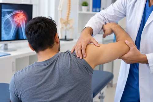
Before reducing the joint, the clinician checks pulses, skin colour and temperature, capillary refill, sensation, movement and nerve function in the affected limb. They document these findings because a dislocation can injure an artery or nerve even when the deformity is obvious. The same checks are repeated after reduction.
X-rays may be taken before reduction when the injury mechanism or examination suggests a fracture. Forcing a fractured joint back can worsen bone and soft-tissue damage. X-rays after reduction confirm that the joint is aligned and show associated bone damage.
| Imaging | What it shows | When it is used |
|---|---|---|
| X-ray | Alignment and many fractures | Initial assessment and after reduction |
| CT | Complex fracture patterns and bone loss | When X-rays do not define the injury |
| MRI | Labral, ligament, tendon and cartilage damage | Persistent symptoms unexplained by X-rays |
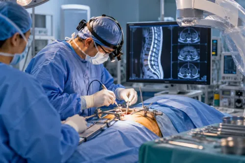
A closed reduction is a controlled manoeuvre that restores the joint without surgery. An open reduction restores the joint in an operating room and is used when closed treatment fails or fracture damage requires an operation. A bone fracture specialist helps decide the safest approach.
Pain control can involve local anaesthetic, intravenous pain medicine, procedural sedation or general anaesthesia, based on the joint, muscle spasm, pain, injury complexity and patient factors. Do not eat or drink before assessment if sedation or surgery may be needed, unless emergency staff instruct you otherwise.
Which joints need hospital care and how fractures change treatment
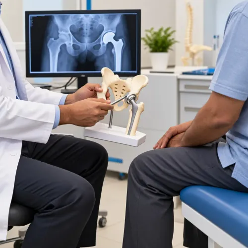
A shoulder, elbow, finger, kneecap or ankle dislocation needs prompt assessment, even when the limb is not severely deformed. A suspected hip dislocation requires urgent hospital care because prolonged displacement can damage the femoral head’s blood supply.
| Joint | Urgency | What may happen |
|---|---|---|
| Shoulder | Prompt assessment | Reduction, imaging and immobilisation |
| Elbow | Prompt assessment | Reduction after checking nerves and circulation |
| Finger | Prompt assessment | Reduction, splinting and fracture assessment |
| Kneecap | Prompt assessment | Reduction, X-ray and bracing |
| Ankle | Prompt assessment | Reduction, splinting and fracture assessment |
| Hip | Emergency hospital care | Urgent reduction, imaging and monitoring |
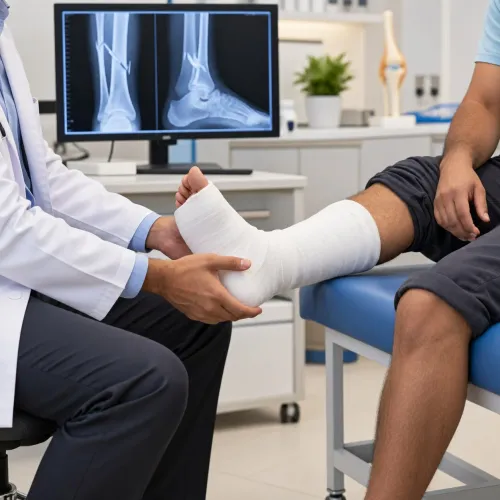
A joint that looks out of place is not automatically a simple dislocation. Bone fractures and joint dislocation can occur together, creating a fracture-dislocation. X-rays help identify it, and treatment may involve casting, fixation, surgery or hospital monitoring rather than straightforward reduction.
Go to an emergency department instead of an outpatient clinic for suspected hip or spine injury, major trauma, an open fracture, uncontrolled bleeding, an absent or weak pulse, severe numbness or weakness, pale or blue cold skin, or a visibly deformed limb after a high-energy accident. Do not attempt reduction yourself.
If you are searching for dislocation for treatment Pimple Saudagar, use this rule: emergency warning signs matter more than proximity. Choose hospital care first when any warning sign is present; seek prompt orthopaedic assessment for other joint dislocations.
What happens after reduction and how recovery is planned
After reduction, the clinician repeats circulation, sensation and movement checks and orders X-rays to confirm alignment and identify bone damage. You may leave with a sling or splint, instructions to elevate the limb and use wrapped cold packs for swelling, and clear limits on lifting or joint movement.
Supervised physiotherapy begins when the joint is stable enough for safe movement. Rehabilitation usually advances from protected movement, to range-of-motion exercises, and then strengthening the muscles that stabilise the joint. Returning to sport or heavy work before strength and control return increases reinjury risk.
Recovery depends on the joint, whether this was a first-time or recurrent injury, an associated fracture, your age, tissue damage, work demands and adherence to rehabilitation—not a fixed timeline. Ask a bone fracture specialist to review the plan when imaging shows a fracture or bone defect.
Seek prompt review for:
- Increasing pain or swelling
- Fever or wound drainage
- Numbness, colour change, or cold fingers or toes
- The joint slipping out again
| Recurrent shoulder finding | Usual direction | Why it matters |
|---|---|---|
| Rehabilitation restores stable control | Continue supervised rehabilitation | Surgery is not automatic |
| Repeated episodes or persistent instability | Discuss arthroscopic or open stabilisation | The shoulder may not stay secure |
| Bone loss, labral or ligament damage, or inability to keep it reduced | Orthopaedic surgical assessment | Repair or stabilisation may be needed |
A shoulder assessment also considers the labrum, ligaments, rotator cuff, bone and nearby nerves before choosing continued rehabilitation or surgery.
How to choose a Pimple Saudagar clinic and prepare for the visit
Choose an orthopedic clinic near me only after screening for danger signs: an open wound, absent or weak pulse, pale, blue or cold skin, numbness, weakness, severe uncontrolled pain, marked deformity or suspected fracture requires emergency-department care, not a nearby outpatient appointment.
| Clinic feature | What to confirm | Why it matters |
|---|---|---|
| Same-day examination | A clinician can assess you today | Delay can worsen swelling, nerve or blood-vessel problems |
| X-ray access | On-site imaging or a direct X-ray referral | Fracture-dislocations cannot be identified by symptoms alone |
| Neurovascular assessment | Pulses, colour, temperature, capillary refill and sensation are documented | Findings must be checked again after reduction |
| Reduction and splinting | Controlled reduction and immediate immobilisation are available | Unplanned attempts can damage bone, cartilage, nerves or arteries |
| Follow-up physiotherapy | Appointments and staged exercises are arranged | Protected movement prevents stiffness and restores strength |
| Hospital pathway | Clear referral for surgery, admission or complex fracture care | Hip, spine and major-trauma injuries need hospital treatment |
Staff will ask when and how the injury occurred, inspect deformity and swelling, record circulation and nerve findings, arrange imaging, discuss reduction or referral, immobilise the joint and set follow-up timing.
Bring prior X-rays or scans, the injury time and mechanism, medicine and allergy details, tetanus history for an open wound, and notes about numbness, colour change or circulation loss. After discharge, plan for repeat appointments, gradual exercises, and restrictions on work or driving.
Latkar Multi-speciality Clinic is an option for orthopaedic assessment and follow-up when outpatient care is suitable; use immediate hospital escalation for the emergency features above.
Related products
 | Tramatology and Fractures surgeon in pune Tramatology and Fractures A bone fracture is a medical condition in which a bone is cracked or broken. It is a break in the continuity of the bone. View product → |
 | Bone Fractures & Joint Dislocation DISLOCATION Bone Fractures & Joint Dislocation Two of the most common bone and joint injuries are fractures and dislocations. |
Frequently asked questions
How should you protect a suspected dislocation before treatment?
Keep the injured joint still in the position found, support the limb with a sling or padding, apply a wrapped cold pack, and avoid trying to push the joint back into place.
What happens during dislocation examination and reduction?
A clinician checks the joint’s shape, movement, circulation and nerve function, then uses imaging such as X-rays to identify dislocation and fractures. Reduction realigns the joint, with pain relief or sedation used when needed.
When does a dislocation need hospital care?
Go to hospital for suspected hip, knee or spine dislocation, an open injury, severe deformity, absent pulse, numbness, cold or discoloured skin, or a suspected fracture-dislocation.
What happens after a joint is put back in place?
Clinicians repeat imaging and neurovascular checks, then may use a sling, brace or immobiliser. Recovery includes follow-up, gradual movement, strengthening and treatment for instability or associated fractures.
How can you prepare for a dislocation treatment visit in Pimple Saudagar?
Bring identification, medical records, medication details, allergy information and previous scans. Note when and how the injury occurred, keep the joint supported, and arrange transport instead of driving yourself.
Keywords